Home » Uncategories » 39+ schlau Bilder Ear Inner Structure / "Ear anatomy, Inner ear, Cochlea Histology, Vestibular ... - Structure of ear comprises three main sections:
Saturday, 14 August 2021
39+ schlau Bilder Ear Inner Structure / "Ear anatomy, Inner ear, Cochlea Histology, Vestibular ... - Structure of ear comprises three main sections:
39+ schlau Bilder Ear Inner Structure / "Ear anatomy, Inner ear, Cochlea Histology, Vestibular ... - Structure of ear comprises three main sections:. The hair cells present in the inner ear of mammals help in sensing the position of the body, in accordance with gravity and maintain the equilibrium. From an anatomical point of view, the ear is conventionally and conveniently divided into three parts: At the base of the canals are the utricle and saccule, each containing a patch of sensory hair cells. Structurally, it consists of two main divisions: Within these cells are tiny particles (otoconia) that help monitor the position of your head in relation to.
In mammals, it consists of the bony labyrinth, a hollow cavity in the temporal bone of the skull with a system of passages comprising two main functional parts: Ear the internal (inner) ear is also called the labyrinth because of its complicated series of canals (figure 4, 5 and 6). The parts of the inner ear are the cochlea, the balance mechanism, the vestibular and the auditory nerve. Inner ear anatomy the outer, middle, and inner ear. The ear canal is the tube that connects the pinna to the inner ear.
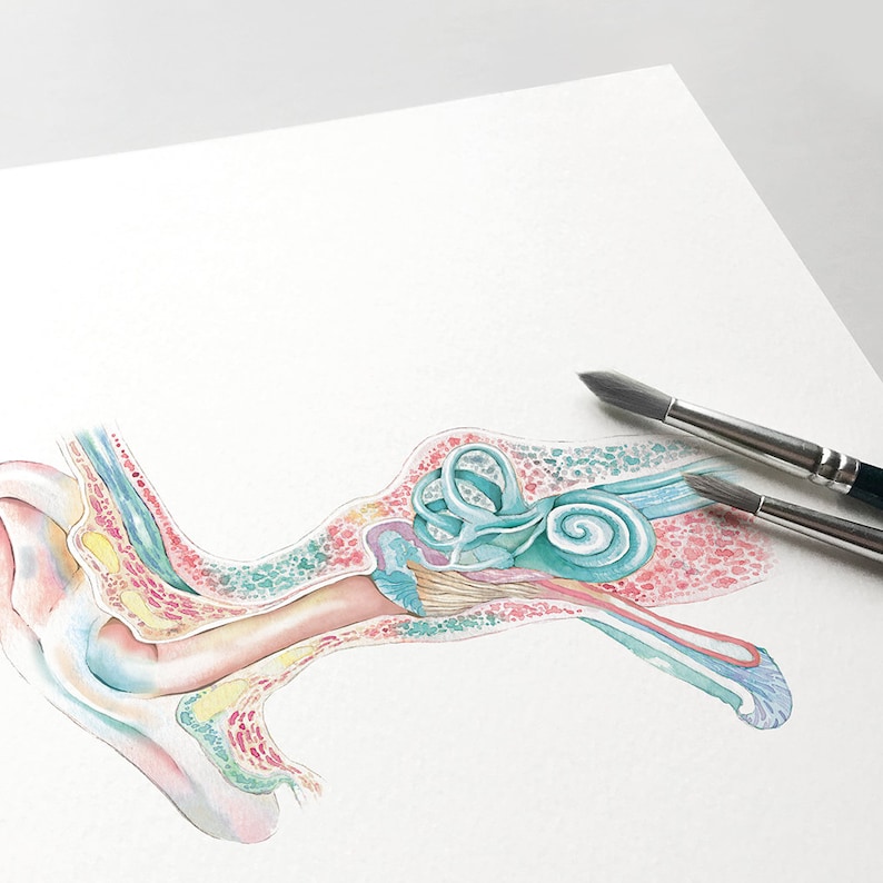
"Ear anatomy, Inner ear, Cochlea Histology, Vestibular ... from ih1.redbubble.net All three parts of the ear are important for detecting sound by working together to move sound from the outer part through the middle and into the inner part of the ear. At the base of the canals are the utricle and saccule, each containing a patch of sensory hair cells. The outer ear is made up of cartilage and skin. The inner ear is also called labyrinth and is composed of two types of labyrinth, i.e. Vintage anatomy print showing a diagram of the inner ear. The oval window is the space where the bone, stapes (or stirrup), makes contact with the inner ear. The middle ear is also called the tympanic cavity or tympanum. The middle ear includes three small bones — the hammer (malleus), anvil (incus) and stirrup (stapes).
There are three different parts to the outer ear;
Bony labyrinth and membranous labyrinth, which in turn are composed of canals and sacs. The outer ear is made up of cartilage and skin. It lies between the middle ear and the internal acoustic meatus, which lie laterally and medially respectively. Each ear consists of three portions: The outer ear, middle ear and inner ear. The outer, middle, and inner ear. The cochlea is the most critical component of the inner ear. The inner ear (internal ear, auris interna) is the innermost part of the vertebrate ear. The middle ear is also called the tympanic cavity or tympanum. The inner ear consists of two functional units: In mammals, it consists of the bony labyrinth, a hollow cavity in the temporal bone of the skull with a system of passages comprising two main functional parts: Inner ear anatomy the outer, middle, and inner ear. Inner ear, also called labyrinth of the ear, part of the ear that contains organs of the senses of hearing and equilibrium.
Bony labyrinth and membranous labyrinth, which in turn are composed of canals and sacs. The parts of the inner ear are the cochlea, the balance mechanism, the vestibular and the auditory nerve. In this article, you will learn more about the inner ear's anatomy. The hair cells present in the inner ear of mammals help in sensing the position of the body, in accordance with gravity and maintain the equilibrium. The inner ear is the innermost part of the ear.
Human ear - Inner ear | Britannica from cdn.britannica.com Bony labyrinth and membranous labyrinth, which in turn are composed of canals and sacs. The inner ear is at the end of the ear tubes. The middle ear is separated from your external ear by the eardrum and connected to the back of your nose and throat by a narrow passageway called the eustachian tube. The ear consists of external, middle, and inner structures. This partition is called the basilar membrane because it serves as. In this article we will discuss about the structure and functions of human ear. The hair cells present in the inner ear of mammals help in sensing the position of the body, in accordance with gravity and maintain the equilibrium. The inner ear consists of tiny bony structures filled with fluid.
In this article we will discuss about the structure and functions of human ear.
What is the inner ear? The human ear is typically divided into three portions: It lies between the middle ear and the internal acoustic meatus, which lie laterally and medially respectively. The bony labyrinth is made up of the cochlea, vestibule, and semicircular canal. The inner ear has two main parts. At the base of the canals are the utricle and saccule, each containing a patch of sensory hair cells. The external ear, middle ear, and inner ear. The inner ear consists of two functional units: This partition is called the basilar membrane because it serves as. This structure helps to give each of us our unique appearance. It consists of two main structural parts one inside the other. The inner ear is also called as labyrinth because of its intricate structure of interconnecting chamber and passage. Structurally, it consists of two main divisions:
The cochlea is the most critical component of the inner ear. At the base of the canals are the utricle and saccule, each containing a patch of sensory hair cells. There are three different parts to the outer ear; This partition is called the basilar membrane because it serves as. Problems with this part of the ear can result in hearing loss and balance issues.
Ear structure Inner Middle Outer Ear Print Biology poster ... from i.etsystatic.com The bony labyrinth, a cavity in the temporal bone, is divided into three sections: Bony labyrinth and membranous labyrinth, which in turn are composed of canals and sacs. The oval window is the space where the bone, stapes (or stirrup), makes contact with the inner ear. The tragus, helix and the lobule. The cochlea is the most critical component of the inner ear. Inner ear anatomy the outer, middle, and inner ear. The inner ear is also called as labyrinth because of its intricate structure of interconnecting chamber and passage. Structure of ear comprises three main sections:
This partition is called the basilar membrane because it serves as.
The ear is made up of three parts: The inner ear is also called labyrinth and is composed of two types of labyrinth, i.e. The outer, middle, and inner ear. Anatomical position and structure the inner ear is located within the petrous part of the temporal bone. The bony labyrinth is made up of the cochlea, vestibule, and semicircular canal. The inner ear (internal ear, auris interna) is the innermost part of the vertebrate ear. In mammals, it consists of the bony labyrinth, a hollow cavity in the temporal bone of the skull with a system of passages comprising two main functional parts: As sound waves travel from the outer to the inner ear, they create waves in the fluid of the inner ear, which in turn moves the tiny hairs in the ear that send sound or movement signals to the brain. Inner ear anatomy the outer, middle, and inner ear. Problems with this part of the ear can result in hearing loss and balance issues. (ii) middle ear and (iii) internal ear. The inner ear is known as the labyrinth, meaning maze; Ears also help to maintain balance.
0 Response to "39+ schlau Bilder Ear Inner Structure / "Ear anatomy, Inner ear, Cochlea Histology, Vestibular ... - Structure of ear comprises three main sections:"


0 Response to "39+ schlau Bilder Ear Inner Structure / "Ear anatomy, Inner ear, Cochlea Histology, Vestibular ... - Structure of ear comprises three main sections:"
Post a Comment